


KPV is a tripeptide: three amino acids, lysine, proline and valine, written in one-letter code as K, P and V. It is the last three amino acids of alpha-melanocyte-stimulating hormone (alpha-MSH), and it is studied in preclinical research for its effects on inflammatory signalling inside cells.
This article explains what the name means, where the sequence comes from, how the Validated Peptides form is built, what laboratories study it for, and how a KPV lot is documented.
Introduction
Most research peptides are tens of amino acids long. KPV is three. That makes it one of the smallest peptides in the catalogue, and its size is a large part of why it is interesting: it is small enough to be carried into cells by a transporter built for the breakdown products of dietary protein.
This article stays with the molecule and the preclinical research around it. Findings described here come from cell lines and animal models, and none of them establish effects in humans.
What KPV Stands For
The name is the sequence. Each amino acid has a one-letter code, and KPV spells out the three in order: K for lysine, P for proline and V for valine. Written in three-letter code, the same sequence is Lys-Pro-Val.
The order matters. Peptides are read from the N-terminus, the end with a free amino group, to the C-terminus, the end with a free carboxyl group. KPV starts with lysine and ends with valine. A peptide with the same three amino acids in a different order would be a different molecule.
The unmodified tripeptide is catalogued in PubChem as MSH (11-13), with the formula C16H30N4O4 and a molecular weight of 342.43 g/mol (PubChem).
Where KPV Comes From: The End of Alpha-MSH
Alpha-MSH is a 13-amino-acid peptide, SYSMEHFRWGKPV in one-letter code. The last three letters are KPV. Researchers number the positions from 1 to 13, which is why KPV also appears in the literature as alpha-MSH(11-13) (Cutuli et al., 2000).
Alpha-MSH has long been studied as a mediator of immunity and inflammation. Its effects on immune cells such as monocytes, macrophages and dendritic cells run largely through the melanocortin-1 receptor, and at the molecular level they involve inhibiting the activation of transcription factors such as NF-kappaB (Luger et al., 2003).
The interest in KPV comes from the finding that this C-terminal fragment keeps part of that activity on its own. The same review reports that KPV was able to bind the MC-1 receptor and modulate the function of antigen-presenting cells (Luger et al., 2003). For research, that turns a 13-amino-acid hormone into a three-amino-acid tool.
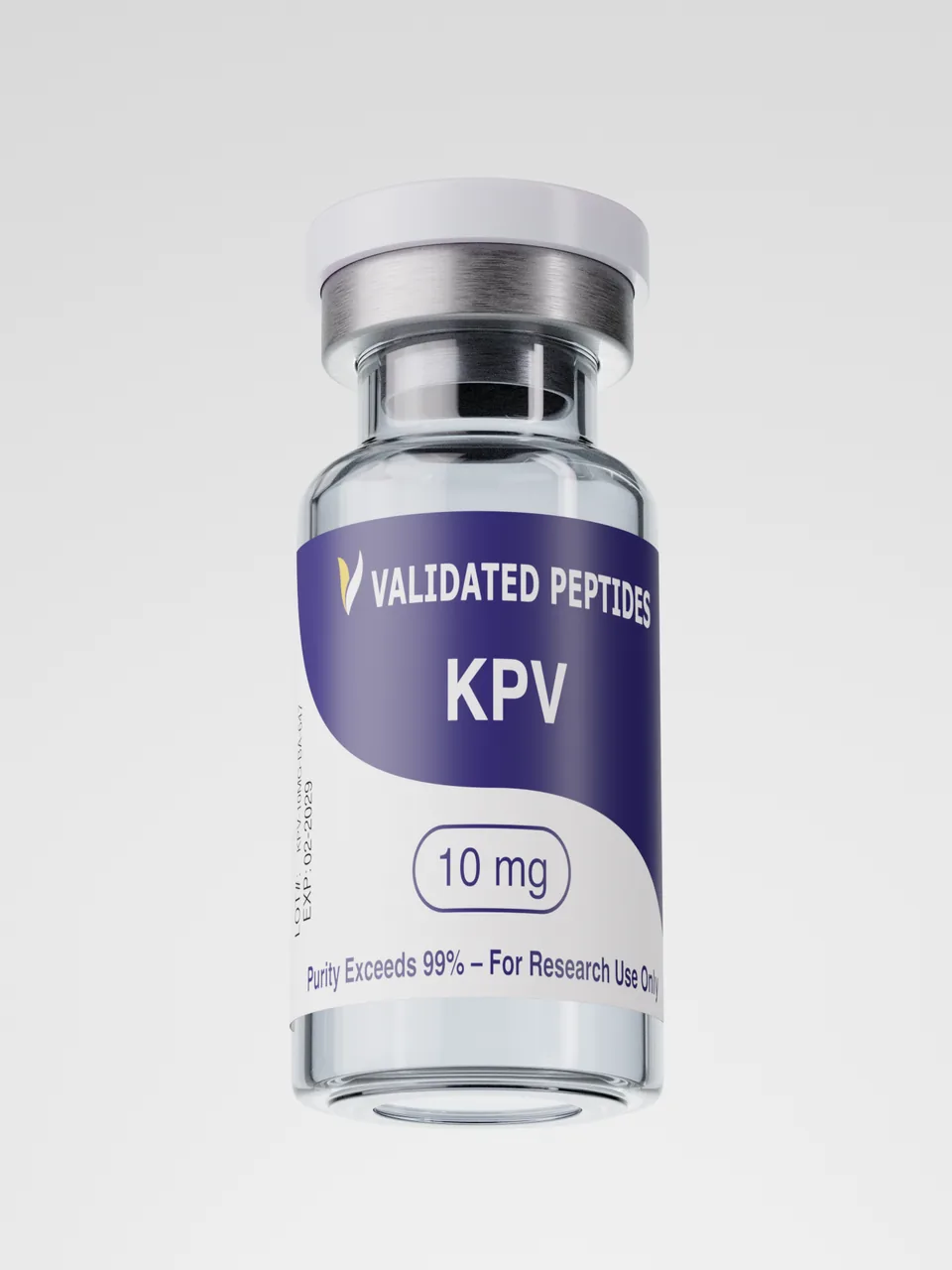
The Validated Peptides Form: Ac-Lys-Pro-Val-NH2
Validated Peptides lists the sequence of its KPV as Ac-Lys-Pro-Val-NH2. The letters on either side of the sequence describe two small changes to the ends of the peptide. Ac stands for an acetyl group on the N-terminus, replacing the free amino group at the lysine end. NH2 stands for an amide on the C-terminus, replacing the free carboxyl group at the valine end.
End-capping of this kind is a common way to make a short peptide more closely resemble the same sequence inside a longer chain, where neither end carries a free charge. It also changes the molecular formula and weight, so a capped KPV and an unmodified KPV are not interchangeable in a study. The form used should be recorded with the lot number, and the identity line on the lot report is what confirms it.
What Researchers Study KPV For
KPV research has concentrated on four questions. All of the work described below is preclinical.
Inflammatory signalling inside the cell
The central body of work looks at NF-kappaB, a protein complex that switches on many inflammatory genes. In a 2008 study, human intestinal epithelial cell lines and human T cells were stimulated with pro-inflammatory cytokines with and without KPV. Nanomolar concentrations of KPV inhibited activation of NF-kappaB and MAP kinase signalling and reduced pro-inflammatory cytokine secretion, measured by a reporter gene, Western blot, RT-PCR and ELISA (Dalmasso et al., 2008).
How KPV gets into cells
The same study asked how such a small peptide acts inside the cell. Using radiolabelled KPV and competition with a known substrate, it found that KPV is transported by PepT1, a di- and tripeptide transporter normally expressed in the small intestine and induced in the colon during inflammation (Dalmasso et al., 2008). That uptake route is why much of the KPV literature centres on intestinal cells.
Animal models of intestinal inflammation
Two 2008 studies extended the cell work into mouse models of colitis, chemically or immunologically induced intestinal inflammation used as laboratory models. Dalmasso and colleagues measured histology and pro-inflammatory cytokine expression (Dalmasso et al., 2008). Kannengiesser and colleagues measured histological change and myeloperoxidase activity, a marker of neutrophil infiltration, and included mice with a non-functional MC-1 receptor (Kannengiesser et al., 2008).
Antimicrobial assays
A separate line of work looked at alpha-MSH peptides, including KPV, against two laboratory pathogens, Staphylococcus aureus and Candida albicans. The peptides inhibited S. aureus colony formation and reduced the viability of C. albicans in vitro, and the authors linked the effect to increased cellular cyclic AMP (Cutuli et al., 2000).
The Open Question: Melanocortin Receptors
One question runs through the KPV literature without a settled answer. Full alpha-MSH acts largely through melanocortin receptors. KPV has been reported to bind the MC-1 receptor (Luger et al., 2003), yet in mice lacking a functional MC-1 receptor, the effects of KPV were still seen, which the authors read as at least partly independent of that receptor (Kannengiesser et al., 2008).
The PepT1 finding offers one explanation: a peptide carried into the cell can act on intracellular signalling directly, without needing a surface receptor (Dalmasso et al., 2008). For study design, it means a KPV experiment can include receptor-blocking or receptor-deficient controls to separate the two routes.
How KPV Studies Are Designed
The published KPV work follows a recognisable pattern, and it is a useful template for reading new studies or planning one.
Concentration ranges
Cell studies have reported effects of KPV at nanomolar concentrations (Dalmasso et al., 2008), and the antimicrobial work on alpha-MSH peptides reported effects across a broad range that reached down to picomolar (Cutuli et al., 2000). With activity reported at such low concentrations, the net peptide content of the vial, not just its label weight, matters when preparing stock solutions.
Endpoints
Signalling work has measured NF-kappaB activation with a reporter gene, protein changes by Western blot, gene expression by real-time RT-PCR and cytokine secretion by ELISA (Dalmasso et al., 2008). Animal model work has added histology and myeloperoxidase activity (Kannengiesser et al., 2008). Choosing endpoints at more than one level, from signalling through to tissue, is what lets a study connect a molecular effect to a model-level one.
Controls
Two control strategies recur. Transport studies used a competing PepT1 substrate to show that uptake runs through that transporter (Dalmasso et al., 2008). Receptor studies used mice with a non-functional MC-1 receptor to test whether the receptor was needed (Kannengiesser et al., 2008). A study that leaves out both cannot say which route an effect came from.
What to Check When Sourcing KPV for Research
Because the capped and uncapped forms are both sold as KPV, the first check is that a listing states which form it is. The sequence written with its end groups, such as Ac-Lys-Pro-Val-NH2, answers that. A listing that says only KPV leaves the form unknown.
From there, the lot report does the work:
- The lot number on the report matches the lot printed on the vial.
- Identity is confirmed by a stated method, so the form in the vial is confirmed rather than assumed.
- Purity is printed against its acceptance limit, so the margin is visible.
- Net peptide content is reported, which matters for a compound studied at nanomolar concentrations.
- Heavy metals, sterility and endotoxin each have their own line and method.
- The report can be checked with the testing laboratory, as set out in the guide to verifying a peptide COA.
Verification
For a peptide this short, identity is the line that matters most. A three-amino-acid sequence leaves little room for error, and a capped and an uncapped form differ only at the ends, so the identity result is what shows the vial holds the intended molecule.
Validated Peptides publishes the current report on the KPV product page and in the Certificate of Analysis library. The current lot:
| Lot | Tested | HPLC purity | Net peptide content | Identity |
|---|---|---|---|---|
| KPV-10MG-BA-647 | August 12, 2026 | 99.34% | 10.54 mg | KPV confirmed by HPLC |
The same lot reports heavy metals not detected by ICP-MS, a pass on a PCR sterility test, and passes on endotoxin and a fentanyl screen. Testing is carried out by an independent laboratory accredited to ISO/IEC 17025.
The guide to reading a peptide COA explains each line of a report, including why purity and net peptide content are reported separately.
Storing KPV in the Lab
KPV ships as a lyophilised (freeze-dried) powder. The published handling guidance is to store it in a cool, dry place away from light, refrigerated at 2 to 8°C, and to avoid repeated freeze-thaw cycles.
When a protocol calls for KPV in solution, it is reconstituted with sterile water or bacteriostatic water. Once dissolved, the solution is stored according to the compound documentation rather than the diluent label.
Where KPV Sits in Peptide Research
KPV is a small, defined tool for one question: what part of the inflammatory signalling activity of alpha-MSH survives in its last three amino acids, and how does it reach the inside of a cell. Its research value rests on that short sequence, on the PepT1 uptake route, and on lot documentation that confirms the exact form in the vial.
KPV is also one of the four peptides in the Klow Blend, alongside GHK-Cu, BPC-157 and TB-500.
Frequently Asked Questions
What is KPV peptide?
KPV is a tripeptide made of lysine, proline and valine. It is the last three amino acids of alpha-melanocyte-stimulating hormone, also written alpha-MSH(11-13). It is studied in preclinical research for its effects on inflammatory signalling in cells. Validated Peptides sells KPV for laboratory research use only, not for human use.
What does KPV stand for?
KPV is the one-letter code for its three amino acids in order: K for lysine, P for proline and V for valine. In three-letter code the same sequence is written Lys-Pro-Val. The Validated Peptides form is listed as Ac-Lys-Pro-Val-NH2, with an acetyl group and an amide capping the two ends.
What does KPV do?
In cell studies, KPV inhibited activation of NF-kappaB and MAP kinase signalling and reduced pro-inflammatory cytokine secretion in intestinal epithelial cells and T cells. It is carried into cells by the PepT1 transporter. These are preclinical findings from cell lines and animal models, and KPV is a research compound, not a medicine.
Is KPV the same as alpha-MSH?
No. Alpha-MSH is a 13-amino-acid peptide, and KPV is only its last three amino acids. KPV keeps part of the inflammatory signalling activity studied for alpha-MSH, but research suggests it may act partly through a different route, by being transported into cells rather than acting only through melanocortin receptors.
How does KPV get into cells?
A 2008 study found that KPV is transported into cells by PepT1, a transporter for di- and tripeptides that is normally expressed in the small intestine and induced in the colon during inflammation. That uptake route is why much of the KPV research has been carried out in intestinal epithelial cell lines.
How should KPV be stored?
KPV should be kept as a lyophilised powder in a cool, dry place away from light, refrigerated at 2 to 8 degrees Celsius, with repeated freeze-thaw cycles avoided. Laboratories reconstitute it with sterile or bacteriostatic water when a protocol calls for a solution, and store that solution according to the compound documentation.
References
- Cutuli M, Cristiani S, Lipton JM, Catania A. Antimicrobial effects of alpha-MSH peptides. Journal of Leukocyte Biology, 2000;67(2):233-239. https://doi.org/10.1002/jlb.67.2.233
- Luger TA, Scholzen TE, Brzoska T, Böhm M. New insights into the functions of alpha-MSH and related peptides in the immune system. Annals of the New York Academy of Sciences, 2003;994:133-140. https://doi.org/10.1111/j.1749-6632.2003.tb03172.x
- Dalmasso G, et al. PepT1-mediated tripeptide KPV uptake reduces intestinal inflammation. Gastroenterology, 2008;134(1):166-178. https://doi.org/10.1053/j.gastro.2007.10.026
- Kannengiesser K, et al. Melanocortin-derived tripeptide KPV has anti-inflammatory potential in murine models of inflammatory bowel disease. Inflammatory Bowel Diseases, 2008;14(3):324-331. https://doi.org/10.1002/ibd.20334
- National Center for Biotechnology Information. PubChem Compound Summary for CID 125672, Msh (11-13). https://pubchem.ncbi.nlm.nih.gov/compound/125672
- International Organization for Standardization / International Electrotechnical Commission. ISO/IEC 17025:2017, General requirements for the competence of testing and calibration laboratories. https://www.iso.org/standard/66912.html